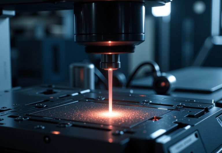
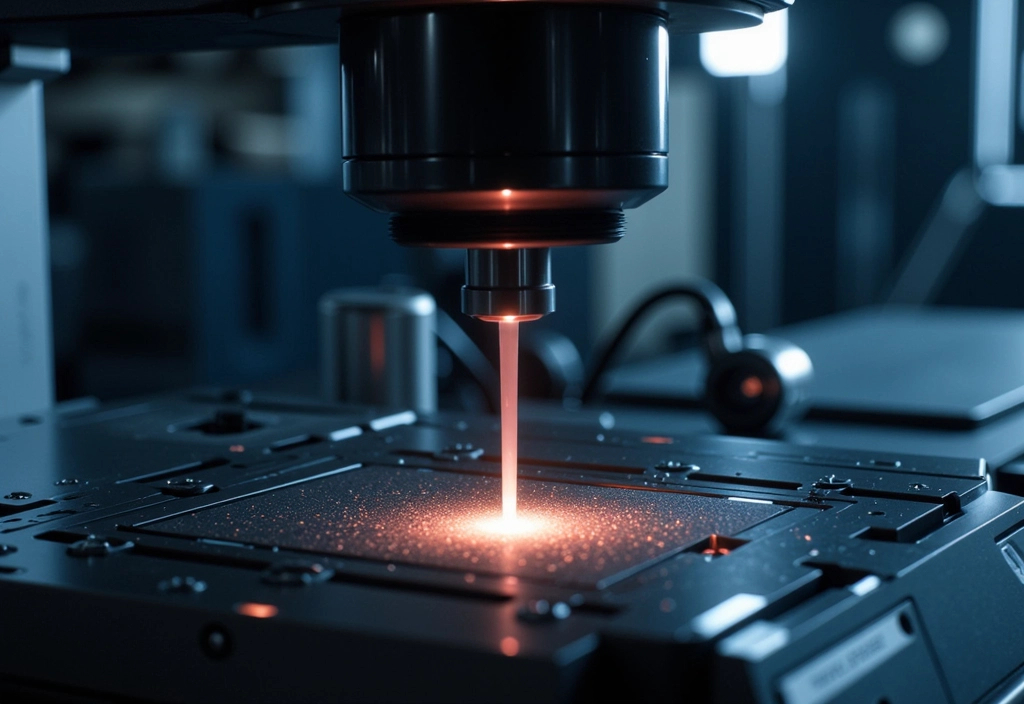
Imagine you’re trying to get a super-clear photo of something really tiny, like an ant, but the ant is running around really fast. That’s kind of like what scientists are doing with electron beams in these plasma accelerators.
- Electron Beams: These are tiny particles moving at almost the speed of light inside a special machine.
- Plasma Accelerator: Think of this like a super-fast racetrack for those electron beams, making them go incredibly fast and in very organized groups.
- 3D Pictures: Scientists wanted to see exactly how these electron groups (or microbunches) look in 3D because this helps them understand and control the beams better.
- Special Camera: They used a special kind of “camera” that can see different colors of light (multispectral imaging) to take detailed pictures of these electron groups.
- Why It’s Cool: By seeing and understanding how these tiny, fast-moving electrons are organized, scientists can make super powerful X-ray lasers. These lasers can then be used to do things like taking really detailed images inside your body (better than current X-rays) or studying materials at an atomic level to create new tech.
In short, it’s like using a super high-tech camera to get a clear picture of something very tiny and fast, which can help us build better tools for science and medicine.
Summary of What Happened:
- What Happened: Scientists successfully developed a method to visualize the 3D structure of electron beams in plasma wakefield accelerators. This technique allows them to see how these beams are organized on a microscopic level.
- Who: A team of researchers working on advanced particle acceleration and imaging technologies.
- Why: Understanding the structure of electron beams is essential for improving X-ray lasers, which have numerous applications in science, medicine, and industry.
How It Works:
- Technique Used: The team used a method called multispectral imaging of coherent optical transition radiation (COTR). This approach allows them to capture images of electron beams as they travel through a plasma accelerator.
- What It Reveals: The method shows the microbunched structure of electron beams, which are crucial for creating highly focused and intense beams used in X-ray lasers.
Benefits to Humanity:
- Medical Imaging: More powerful and precise X-ray lasers could revolutionize medical imaging, making it possible to see finer details in the human body.
- Materials Science: These lasers could also be used to study materials at an atomic level, leading to new advancements in technology and materials.
- Compact Devices: The technology promises to make X-ray lasers more compact and accessible, broadening their use across various fields.
When It Will Be Available:
- Current Stage: The technology is still in the research phase. It shows great promise, but further development and testing are needed before it can be widely used.
- Future Outlook: With ongoing research, we might see practical applications of this technology in the next few years, especially in scientific research and high-tech industries.
Disclaimer: This content was simplified and condensed using AI technology to enhance readability and brevity.
Article derived from : LaBerge, M., Bowers, B., Chang, Y., Cabadağ, J. C., Debus, A., Hannasch, A., Pausch, R., Schöbel, S., Tiebel, J., Ufer, P., Willmann, A., Zarini, O., Zgadzaj, R., Lumpkin, A. H., Schramm, U., Irman, A., & Downer, M. C. (2024). Revealing the three-dimensional structure of microbunched plasma-wakefield-accelerated electron beams. Nature Photonics. https://doi.org/10.1038/s41566-024-01475-2


